Positron emission tomography (PET scan)
Medically reviewed by Drugs.com. Last updated on Feb 2, 2023.
What is Positron emission tomography (PET scan)?
A positron emission tomography, or PET, scan is an imaging technique that uses radioactive tracers attached to sugar molecules. The scan can detect changes in the body's metabolism and chemical activities. A PET scan is often done in conjunction with a CT scan. The combination provides a color-coded image of the body's structure and function.
During a PET scan, the sugar molecules with the attached radioactive tracers are injected into a vein. Once the substance enters the body, it travels through the bloodstream to the body's organs. Areas in the body that have a higher rate of metabolism use more sugar. So these areas pick up the sugar molecules more readily than normal.
The PET scanner uses computers to interpret the amount of tracer uptake in different parts of the body to create the color-coded images. Conditions that typically have higher metabolic rates include cancers, infections and neurological diseases.
A PET scan is painless, except for a mild skin prick when the substance is injected.
What it's used for
A PET scan may be used to evaluate people with the following illnesses:
- Cancer — PET scans can be used to detect cancerous tumors, to determine how much cancer has spread) and to determine how well cancer treatment is working. They are used most often in patients with brain cancer, colorectal cancer, lymphoma, melanoma and lung cancer.
- Brain diseases — PET scans can be used to evaluate neurological illnesses, such as epilepsy, and Alzheimer's disease and other dementias.
- Cardiac illnesses — PET scans can be used to evaluate how well the heart muscle is functioning in patients with coronary artery disease or cardiomyopathy.
- Infected organs and abscesses — PET scans can be used to help diagnose unexplained fevers due to hidden infections.
PET scans also are used for research in other areas, including drug addiction, psychiatric illnesses and stroke. Medical specialists are just beginning to discover how PET scans can be used to evaluate a wide range of patients. New uses are being found every year. Because some types of PET scans still are considered experimental by some medical insurers, check with your doctor and your medical insurer before your scan to verify your coverage for this procedure.
Preparation
Because a PET scan involves radioactivity, tell your doctor if you are pregnant or if there is a possibility that you might be pregnant. Tell your doctor if you think you will be unable to lie very still for 30 minutes to two hours during the PET scan.
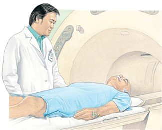
How it's done
A PET scan usually is done as an outpatient test in a major medical center that has a small cyclotron, very advanced nuclear medicine equipment used to make the PET tracer.
The PET scanner is a ring-shaped apparatus with an attached table. You will lie on the scanning table, and the table will slide slowly through the opening in the scanner ring. One or two scans might be taken before the tracer is administered. After this initial scanning, the tracer will be injected into one of your veins, usually in your arm. Additional scans will be taken while the tracer is in your body.
During the scanning procedure, you must lie very still. The scanning table will glide you through the PET scanner, so you won't need to move. If your head is being scanned, special cushions may be placed against your head to hold it in place.
Plan to spend about three hours to complete the test. Afterward, you can go home and resume your normal activities.
|
|
Follow-up
Ask personnel at the scan facility about when you should call your doctor for the official scan report.
Risks
The radioactive tracers used in PET scans are considered to be safe, and because they are short-lived, they are quickly cleared from the body.
When to call a professional
If the tracer was injected, call your doctor if you have pain, redness or swelling at the injection site.
Additional info
Society of Nuclear Medicine and Molecular Imaging (SNMMI)
https://www.snmmi.org/
National Library of Medicine (NLM)
https://www.nlm.nih.gov/
Further information
Always consult your healthcare provider to ensure the information displayed on this page applies to your personal circumstances.